

The hippocampus is a critical brain region that plays essential roles in various cognitive functions, such as memory formation, retrieval, learning, decision-making, and the regulation of emotional responses. Despite extensive research into its structure and organizational function, a complete understanding of the cell types present in this area and their connectivity with other neurons remains unresolved.
Over recent years, methods for studying these cellular subpopulations, gene expressions within them, and their connectivity have greatly advanced. Two prominent techniques include spatially resolved transcriptomics (SRT), which preserves the physical arrangement of cells while measuring gene expression, and single-nucleus RNA-sequencing (snRNA-seq), which identifies differences between individual cell nuclei by examining their RNA molecules.
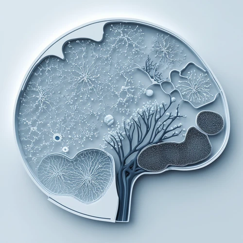
Researchers from Johns Hopkins Bloomberg School of Public Health, Lieber Institute for Brain Development, and Johns Hopkins School of Medicine have utilized both SRT and snRNA-seq to analyze hippocampal tissue from adult human donors. Their findings are detailed in a recent publication in Nature Neuroscience, where they introduce an extensive molecular atlas mapping cell subtypes and their organization.
Nature Neuroscience"The specialized functions of different hippocampus cell types, evident through unique morphology, physiology, and connectivity patterns, are spatially organized," wrote Jaqueline R. Thompson, Erik D. Nelson and co-authors in their paper. "Retaining the cytoarchitectural organization requires advanced molecular profiling approaches. We generated SRT and snRNA-seq data from anterior regions of the human hippocampus in ten neurotypical adult donors."
Nature NeuroscienceThe team studied hippocampal tissue collected from brains with no reported anomalies belonging to 10 individuals, and obtained combined SRT and RNA-seq datasets through computational methodologies.
"We integrated these datasets using non-negative matrix factorization (NMF) and label transfer by identifying gene expression trends within the snRNA-seq data and inferring analogous patterns in the SRT dataset," wrote Thompson, Nelson, and colleagues. "These patterns capture transcriptomic variations among neuronal types and demonstrate spatial organization of synaptic specializations for excitatory and inhibitory neurons."
Through their detailed examination, researchers were able to pinpoint the locations of various cell subtypes within the hippocampus and compare these findings with data from mouse models.
"Using NMF and label transfer techniques on rodent datasets, we uncovered potential patterns of activity-dependent transcription and circuit connections in human SRT data," wrote Thompson, Nelson, et al. "Finally, we characterized spatial arrangements of NMF patterns linked to pyramidal neurons, revealing distinct snRNA-seq clusters unique to the retrohippocampus, subiculum, and presubiculum regions."
The molecular atlas developed by the researchers is accessible online via an interactive web application and includes raw data that could be invaluable for future neuroscience or medical research focusing on the hippocampus.
This article was meticulously crafted by our author Ingrid Fadelli, edited by Gaby Clark, and reviewed and fact-checked by Robert Egan. We depend on readers like you to keep independent science journalism alive! If this article is important to you, please consider making a donation—especially if it's monthly. As gratitude, you'll receive an ad-free account.
Ad-Free