

A collaborative study between UT Health San Antonio and Stanford University researchers has significantly advanced our comprehension of how the body perceives different sensations, including pain, itch, and touch.
When a person experiences pain or any external stimulus, primary sensory neurons translate these signals into electrical impulses to prompt an appropriate response. While this process was known, its specifics—how different types of sensation are distinguished—remained unclear, until now.
Researchers at The University of Texas Health Science Center at San Antonio (UT Health San Antonio) and Stanford have created a novel in vivo imaging system that reveals, in real time, how neurons activate to represent various sensations. Their research is featured in the July 2025 edition of Nature Communications.
The study marks a substantial leap forward in understanding sensation processing within the nervous system and paves the way for new treatments against pain and sensory disorders.
Primary sensory neurons handle somatosensation—the conversion of stimuli like touch, pressure, pain, itch, and proprioception into brain signals. Despite extensive research over decades, how these neurons differentiate sensations remained a mystery.
Nature Communications"When there's a stimulus such as injury, we instantly sense pain simultaneously with touch. But knowing what makes us perceive one versus the other has been elusive," said Yu Shin Kim, Ph.D., study co-author and associate professor in the Department of Oral and Maxillofacial Surgery at UT Health San Antonio. "This basic mechanism has been a long-standing question in neuroscience."
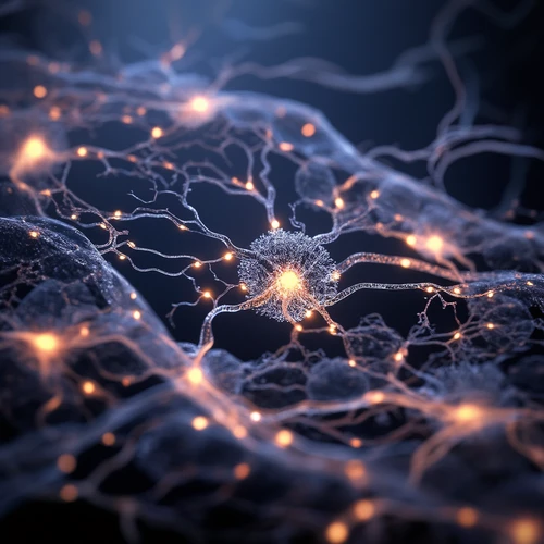
The team's work revolves around ASAP4.4-Kv, a genetically-encoded voltage sensor they developed. This positively-tuned sensor exhibits increased fluorescence when neurons depolarize, allowing researchers to observe neuron communication in real time within mouse models. The imaging system uses sensors that light up upon neuron activation, enabling scientists to pinpoint which neurons are firing and their responses to various sensations under a powerful microscope.
"With this technology, we've made it possible to see electrical signals from these neurons and link them to different types of sensation for the first time," Kim said.
The imaging technique verified an existing hypothesis that sensory neurons communicate electrically after inflammation or nerve injury.
Nature Communications"Post-inflammation or injury, we've confirmed these neurons do indeed communicate electrically. They transmit extremely quick signals so each neuron can convey its activity to another," Kim said. "This theory is now visualized and verified thanks to our work."
Compared to traditional electrophysiological recordings that are time-consuming and invasive, this technique offers many advantages. It allows researchers to observe sensory neuron subtypes continuously.
The applications of this technology include investigating chronic pain, inflammation, itch, temporomandibular joint (TMJ) pain, migraine, and other somatosensory conditions.
"The ability to observe these reactions was absent before; now we have the means," Kim said.